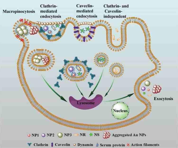
The size, shape and protein corona of gold nanoparticles play key roles in cellular uptake and removal mechanisms. 15 nm nanoparticles (NP1), 45 nm nanoparticles (NP2), and rod-shaped nanoparticles (NR) enter cells via a receptor-mediated endocytosis (RME) pathway. Stellar nanoparticles (NS) are not only mediated by clathrin, but also through the endocytosis pathway mediated by lumenin. However, due to its large size, 80 nm nanoparticles (NP3) mainly enter cells through the macocytosis pathway. In addition, the presence of protein corona can alter the absorption mechanism of Au NPs. The endocytosis pathway of NP1, NP2 and NS was changed from RME pathway to macocytosis pathway, and NR was changed from RME pathway to Clathrin and Caveolin independent pathway under non-FBS inclusion. Five Au NPs, both coated and uncoated, were released via the lysosomal exocrine pathway. The size and shape of the protein crown affected the exocytosis rate and exocytosis volume, but did not change the mechanism of exocytosis. Systematic study of the mechanism of endocytosis and exocytosis of gold nanoparticles of different sizes and shapes is helpful for the toxicological evaluation of gold nanoparticles and the application of nano-drugs.
We studied the absorption, removal and mechanism of five Au NPs. We demonstrate that size, shape, and protein crowns play key roles in the cellular uptake and removal mechanisms of Au NPs. The absorption mechanisms of these NPs are different. The uptake of NP1, NP2 and NR is a clathrin receptor-mediated endocytosis pathway. NS is not only a clathrin-mediated endocytosis pathway, but also a cave-protein mediated endocytosis pathway. NP3 aggregates mainly through macropinocytosis due to its large volume. Protein crowns reduce the aggregation of NPs, resulting in reduced cell intake. We also found that protein crowns affect the mechanism of endocytosis. The endocytosis pathway of non-FBS coated NP1, NP2 and NS was transformed from RME pathway to macytosis pathway. Size, shape and protein crown had no effect on the mechanism of exocytosis. Five kinds of Au NPs are released through lysosomal exocytosis. The exocytosis rate and the amount of exocytosis are related to each other. Smaller NPs seem to escape at a faster rate. This research is helpful to understand the endocytosis and exocytosis mechanism of gold NPs, and provides guidance for the application of gold NPs in the field of biomedicine.
https://onlinelibrary.wiley.com/doi/full/10.1002/smll.201801451